Mangino Research Lab
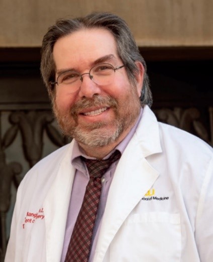
Research Lab of Dr. Martin J. Mangino
Publications
Click here for a list of publications
Research Goals
- To understand the molecular mechanisms of ischemia reperfusion injury
- To translate new treatments for clinical manifestations of reperfusion injury such as organ preservation, shock, critical illness, and acute organ failure
- To educate and train new medical research scientists in basic and translational science.
Lab Members
_copy.png)
Caitlin Archambault, LVT
Lab Manager
_copy.png)
Caitlin Archambault, LVT
Lab Manager
Surgery
_copy.png)
Ru Li, MD, PhD
Senior Scientist
_copy.png)
Ru Li, MD, PhD
Senior Scientist
Surgery
_copy.png)
Loren Liebrecht, MD
Post-Doctoral Scholar
_copy.png)
Loren Liebrecht, MD
Post-Doctoral Scholar
Surgery
_copy.png)
Jerry Maitland
PhD Candidate
_copy.png)
Jerry Maitland
PhD Candidate
Department of Pharmacy
_copy.png)
Charles Payne
MS Student
_copy.png)
Charles Payne
MS Student
Department of Physiology and Biophysics
_copy.png)
Priyanshi Parikh
MS Student
_copy.png)
Priyanshi Parikh
MS Student
Department of Physiology and Biophysics
Research Program
Research Focus
Maintaining adequate cell and tissue bioenergetic function through mitochondrial oxidative metabolism is the basis of normal cell function in health and the basis for almost half of medical diseases. The balance between the delivery of oxygen to cells and tissues and the utilization of the energy needs of the tissue is essential for normal function. Imbalances in oxygen delivery to demand in mitochondria is the basis for both ischemic disorders and the blueprint for treating the problem. As cell bioenergetic begins to fail because of ischemia, then secondary, tertiary, and further downstream mechanism serve to complicate and exacerbate the condition. Our strategy is to find mechanism of ischemic injury at the basic science level that affect the root cause of the problem. which is the delivery of oxygen to tissue, the utilization of oxygen, and the sensitivity of the tissue to loss of ATP synthesis to mitigate the root cause of ischemic injury, thereby short-circuiting and rendering moot the downstream mechanisms. The ultimate goal is to translate effective clinical treatments for ischemic disorders from our mechanistic paradigms developed in the lab and to deploy these treatments through commercialization strategies for broad use to patients to extend life and relieve human suffering.
Shock, trauma, and critical illness
My lab studies mechanisms of acute resuscitation injury in trauma and critical illness. Mechanisms of global reperfusion injury after resuscitation from severe shock leverage new concepts learned for the development of new intravenous low volume resuscitation solutions. New mechanisms of loss of local capillary blood flow after severe shock that involve the natural loss of ATP-dependent cell volume control mechanisms have been studied in detail. New research tools developed to selectively “knock-out” these osmotic shifts have conclusively demonstrated the significance of this local mechanism in shock and reperfusion injury in many others settings such as acute kidney injury, myocardial infarction, splanchnic ischemia, sepsis, cardiac arrest and TBI. We have proposed a paradigm shift to describe the mechanism causing reperfusion injury and are trying to promote that shift because the old paradigms don’t work. This new understanding and the new tool used to test these hypothesis worked so well in the lab that they are being developed, commercialized, and tested in human trials of severe hemorrhagic shock. This led to the launch of Perfusion Medical, Inc. to make these new stable and effective research tools available to trauma patients and patients in the ICU with critical illness and sepsis. Other advancements in shock include the importance of metabolic changes that occur in shock, trauma, and critical illness that lead to aberrations in local nitric oxide metabolism and altered oxygen delivery to tissues after reperfusion. Finally, new synthetic blood substitutes and stable bridges to blood transfusion are being developed for the US Department of Defense to treat severe shock, trauma, and blood loss in the austere pre-hospital environment in military and civilian medicine. These components drastically improve the microcirculation to improve efficiency of oxygen transfer after resuscitation with or without blood components, the replacement of oxygen carriers with biosynthetic stable alternatives, and the recapitulation of coagulation and platelet function with stabilized and dried plasma components, and recombinant proteins. Restoration of oxygen transfer to ischemic organs to pay back oxygen debt in the microcirculation after shock as rapidly as possible is highly effective, lifesaving, and is needed in the austere pre-hospital and early hospital space for trauma patients.
Organ preservation for transplantation
Organs recovered from cadaver donors for transplantation into recipients with end stage organ failure undergo a period where the oxygen delivery is interrupted after it is recovered. Unless something is done between the time of recovery and the time of reperfusion at transplantation, irreparable ischemia will destroy the function of the grafts. The science of maintaining donor organ function outside of the donor is called organ preservation and the reperfusion injury that occurs after transplantation is organ preservation injury. My laboratory has been working on this problem for over 35 years where significant mechanisms of injury and principles guiding organ preservation have been developed. These principles led to the development of modern organ preservation solutions and techniques. However, advancements in organ preservation have not been realized in almost 30 years since these discoveries because we have failed to continue understanding more basic root molecular mechanisms of injury in these organs and tissues. That basic work continues in the lab and discoveries in these basic models often informs our treatment strategies for other clinical manifestations of reperfusion injury in shock, MI, compartment syndrome AKI, etc. Organ preservation projects in the lab focus on the cytoskeletal signaling changes that have been shown to be very important in liver preservation injury where specific signaling inputs from lipid mediators and mediator hormones (Lyso-phosphatidic acid and sphingosine-1-phosphate) maintain sub-lamellar cytoskeletal proteins (moesin, radixin, and ezrin) in active binding configurations necessary for cell membrane and mitochondrial function. Restoration of these lipid paracrine hormones and developing stable LPA and S1P receptor agonists are being explored to maintain cytoskeletal integrity during cold storage preservation and machine perfusion preservation. Mutant animals and cells expressing these mutant cytoskeletal proteins are used to test these hypothesis. Other projects involve the reanimation of organs recovered from patients in the emergency department that die from failed resuscitation. These donors and their organs are currently not usable for organ transplantation because of the rapid preservation injury incurred because of their cause of death. We are using mid-thermic machine perfusion strategies in the lab and cardiopulmonary bypass strategies in the ED to support these organs after death. Preliminary experiments have shown it is possible to transfect genes into these isolate livers and express or de-express genes of interest that could be helpful for restoring organ function. Other approaches in reanimation include repairing defects in mitochondrial complex-1 electron transfer to restore aerobic ATP synthesis and altering differential gene expression by influencing epithelial to mesenchymal transition (EMT) that is induced in livers and kidneys with severe preservation injury. We have identified EMT as a basic molecular mechanism of ischemic cholangiopathy in liver grafts with preservation injury. Liver cholangiocyte EMT-induced ischemic cholangiopathyis a delayed preservation injury seen in livers transplanted with preservation injury suffered at donor organ recovery. Preventing and treating ischemic cholangiopathy, as we are doing now in the lab using novel pharmacological tools targeting EMT signaling, will allow the use of these damaged livers for clinical transplantation. This will drastically increase the donor pool and remove dying patients from the liver wait list. The lab has a dedicated space for organ reanimation of human donor organs recovered in the VCU Emergency Department to translate these findings to clinical therapeutic options for our transplant service.
Cytoskeletal system in ischemia
The lab continues to conduct basic science experiments in cell culture models to understand how changes in the cytoskeletal system cause ischemic injury in preserved organs and in any condition where ischemia occurs. Targets of interest include signaling that controls activation and deactivation of ERM proteins, how ERM proteins may bind to the outer mitochondrial membrane to alter the mitochondrial permeability transition pore from opening, and how “treadmilling” of actin and tubulin polymerization is involved in reperfusion injury. Finally, the link between degraded bioenergetics in ischemia and cytoskeletal changes is also studied. It is often easier to prevent cytoskeletal degradation by maintaining cellular ATP levels during and after organ recovery or restoring ATP levels quickly after reperfusion than to fix degraded cytoskeletal systems.
Teaching and Mentoring
Mentoring and teaching is an important component of the Mangino Lab. We train young folks to become biomedical research scientists that can develop innovative ideas, test hypothesis in the basic science lab in cell and molecular models, advance the project in higher order models, pre-clinical testing, and clinical trials in patients. In fact, the lab moto is curing disease from molecule to bedside, which is what we offer our students. An important new component of the lab is to teach students and faculty skills to bridge the gap between academic testing in pre-clinical models to testing in humans. This often requires commercialization skills to incentivize the funding and de-risking of the final product through FDA compliance so it can be used safely in human trials. These business related skills are often missing in academic research labs and are a major reason great ideas developed in academic labs are lost forever. In the lab, we teach students critical thinking and problem solving, data analysis, the scientific and epistemological philosophy of thinking, and hands on training in modern scientific methods, including molecular biology, physiology, pharmacology, immunology, surgery, and clinical trials skills. Fundraising is critical since a lab goes nowhere without funding. Students are taught how to prepare grants to compete at federal agencies like NIH, DoD, NSF, and private sources. I have trained over 100 medical and surgical residents, undergraduate students, PhD and MS students, medical students, and junior faculty in the lab over the last 38 years.
